Complexity of severe asthma

Inflammation in asthma is complex and heterogeneous, which makes it challenging to manage.1–4

Severe asthma is characterized by complex, heterogeneous, and dynamic airway inflammation1–4

  • Activation of the airway epithelium, following exposure to environmental triggers, results in epithelial cytokine release1,5,6
  • This release of epithelial cytokines can lead to initiation of multiple downstream inflammatory pathways, including Type 2 (T2) inflammation (allergic and/or eosinophilic) and beyond T2 inflammation (structural changes to the airway)1,6–9
  • Patients may display overlapping, dynamic inflammatory pathways, which can change over time owing to different circumstances1–4,9; many patients are thought to have multiple drivers of disease2
  • Biomarkers are objective, measurable diagnostic tools that can change over time or with treatment and can help to identify phenotypes and endotypes of severe asthma. Biomarkers serve as valuable tools to support clinical decision making10

Improved understanding of the key inflammatory pathways and mechanisms that underpin epithelial-driven diseases will continue to drive research and development of new treatment approaches, with the goal of finding ways to reduce epithelial-driven inflammation and restore epithelial health, lower disease activity, alter disease progression and ultimately achieve clinical remission in asthma4,6,11–13

References
1. Busse WW. Allergol Int. 2019;68:158–166; 2. Tran TN, et al. Ann Allergy Asthma Immunol. 2016;116:37–42; 3. Price D, Canonica GW. World Allergy Organ J. 2020;13:100380; 4. Russell RJ, et al. Eur Respir J. 2024;63:2301397; 5. Lambrecht BN, Hammad H. Nat Med. 2012;18:684–692; 6. Gauvreau GM, et al. Expert Opin Ther Targets. 2020;24:777–792; 7. Cao L, et al. Exp Lung Res. 2018;44:288–301; 8. Wu J, et al. Cell Biochem Funct. 2013;31:496–503; 9. Kuruvilla ME, et al. Clin Rev Allergy Immunol. 2019;56:219–233; 10. Carr TF, Kraft M. Ann Allergy Asthma Immunol. 2018;121:414–420; 11. Kaur R, Chupp G. J Allergy Clin Immunol. 2019;144:1–12; 12. Agache I, et al. Allergy. 2012;67:835–846; 13. Brightling CE, et al. Eur Respir Rev. 2024;33:240221.

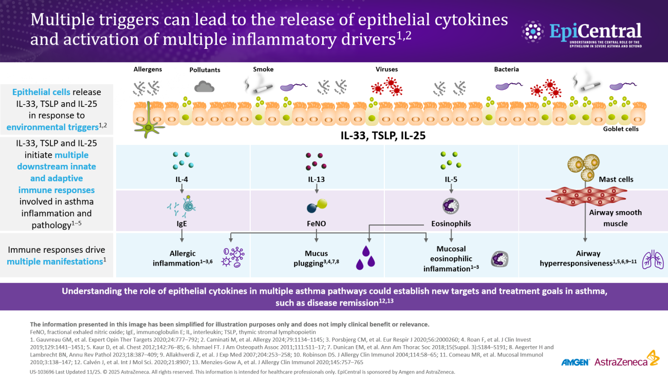
Inflammation in severe asthma

Asthma-associated inflammation is complex and heterogeneous,1–4 and numerous cell types, mediators, and downstream immune pathways are involved.1–6 Multiple inflammatory endotypes have been characterized, including allergic and eosinophilic inflammation.6

Video: Watch Professor Christopher Brightling introduce the complexity of severe asthma ​ (04:09)

Image
US EpiCentral_Module 2_inflammatory cascade_hero asset

Multiple triggers can lead to the release of epithelial cytokines and activation of multiple inflammatory drivers.7–10 Patients with severe asthma have an average of eight different triggers that exacerbate asthma symptoms via the release of epithelial cytokines, such as thymic stromal lymphopoietin (TSLP), which can activate multiple inflammatory drivers and promote airway hyperresponsiveness.7–10

Download resource

For a deep dive on the inflammatory pathways that underpin the complexity of inflammation in severe asthma, please listen to Dr. Jonathan Corren here from 06:45 min.

Overlapping and changing inflammatory pathways in severe asthma

Activation of the airway epithelium by exposure to environmental triggers results in the production of epithelial cytokines and leads to a cascade of events that result in airway inflammation and the clinical manifestations of asthma.11,12 This airway inflammation drives changes in asthma pathophysiology and leads to airway hyperresponsiveness and airflow obstruction.1

The production of epithelial cytokines leads to multiple downstream immune pathways in patients with severe asthma, including: 

  • T2 inflammation, typically marked by allergic and/or eosinophilic inflammation1,12
  • Mechanisms beyond T2 inflammation, characterized by neutrophilic or paucigranulocytic inflammation, airway hyperresponsiveness, airway remodeling, or microbial dysbiosis, which may exist together with T2 inflammation1,12–14

While many patients have T2 disease,1 a sizable group has mechanisms that are beyond T2 disease.2  

Video: Watch Professor Ian Pavord discuss the asthma heterogeneity and the dynamic nature of airway inflammation in asthma (00:47)

Patients may have multiple drivers of inflammation, as seen in the International Severe Asthma Registry (ISAR), where 59% of patients had two or more elevated biomarkers, characterized by elevated immunoglobulin E (IgE), fractional exhaled nitric oxide (FeNO), and/or blood eosinophils of ≥300 cells/µL.15 Therefore, it is likely that many patients with severe asthma have a mixture of pathways driving their disease.1,2

The wide spectrum and overlap of downstream pathways in severe asthma make diagnosis and treatment challenging.1,2

Biomarkers in severe asthma 

Diagnostic biomarkers can help identify endotypes of severe asthma and help in the selection of an appropriate treatment. Various biomarkers of T2-mediated inflammation, including specific blood IgE, blood or sputum eosinophils, and FeNO, are available to clinicians,1,16,17 and these, among others, can be used in clinical practice for phenotyping of severe asthma.16

  • Allergen-specific blood IgE levels, measured via blood or skin prick testing, are higher in patients with allergic asthma compared with healthy individuals and can be used as a surrogate measure for atopy16,18
  • Blood eosinophils and sputum eosinophils are surrogate markers of T2 inflammation and the T2-inflammatory cytokine, interleukin (IL)-5, which is required for eosinophil activation and survival.17 These are useful biomarkers as patients with higher eosinophil counts are prone to experiencing severe disease and poorer asthma outcomes than patients with lower eosinophil counts19
  • FeNO is a biomarker of airway epithelial cell exposure to IL-13 and IL-4.17,20 These cytokines upregulate inducible nitric oxide synthase (iNOS) in the airway epithelium and result in increased nitric oxide production.17 A high FeNO measurement correlates with airway eosinophilia in asthma and indicates increased airway T2 inflammation17

Image
Case study

Biomarker assessment in a 54-year-old man with severe asthma.

Download resource

It is important to note that biomarker levels may be affected by various factors.18 Biomarkers of T2 inflammation are often suppressed by inhaled corticosteroids and oral corticosteroids (OCS); therefore, eosinophils and FeNO assessments are encouraged before commencing a short course or maintenance OCS, or while the patient is on the lowest possible OCS dose.18

Some gaps exist in the clinical predictive value of existing biomarkers due to the challenge of identifying a single predominant endotype of severe asthma.6 Furthermore, there are currently no readily available biomarkers in clinical practice that identify T2-independent asthma.20–22 For now, biomarkers need to be interpreted alongside symptoms and lung function, and treatable features of asthma, such as airway hyperresponsiveness.23

Video: Watch Professor Christopher Brightling ​explain the use of biomarkers in airway inflammation and their predictive value (07:03)

Find out more about the EpiCreator – Professor Christopher Brightling.

RELATED RESOURCES

Like this page? We have a host of other resources available in the ‘Scientific and Resource Library’ where you can find out more.

References

1. Busse WW. Allergol Int. 2019;68:158–166; 2. Tran TN, et al. Ann Allergy Asthma Immunol. 2016;116:37–42; 3. Price D, Canonica GW. World Allergy Organ J. 2020;13:100380 (Abstract OC35); 4. Kupczyk M, et al. Allergy. 2014;69:1198–1204; 5. Barnes PJ. Pathophysiology of asthma. In: European Respiratory Society Monograph. 2003 p. 84–113; 6. Gauvreau GM, et al. Expert Opin Ther Targets. 2020;24:777–792; 7. Chipps BE, et al. Ann Allergy Asthma Immunol. 2023;130:784–790.e5; 8. Lambrecht BN, et al. Immunity. 2019;50:975–991; 9. Lambrecht BN, Hammad H. Nat Immunol. 2015;16:45–56; 10. Bartemes KR, Kita H. Clin Immunol. 2012;143:222–235; 11. Lambrecht BN, Hammad H. Nat Med. 2012;18:684–692; 12. Russell RJ, et al. Eur Respir J. 2024;63:2301397; 13. Cao L, et al. Exp Lung Res. 2018;44:288–301; 14. Wu J, et al. Cell Biochem Funct. 2013;31:496–503; 15. Denton E, et al. J Allergy Clin Immunol Pract. 2021;9:2680–2688.e7; 16. Carr TF, Kraft M. Ann Allergy Asthma Immunol. 2018;121:414–420; 17. Peters MC, et al. Curr Allergy Asthma Rep. 2016;16:71; 18. Global Initiative for Asthma (GINA). Global strategy for asthma management and prevention. 2025. Accessed January 2026.  https://ginasthma.org/; 19. Kostikas K, et al. Curr Drug Targets. 2018;19:1882–1896; 20. Kuruvilla ME, et al. Clin Rev Allergy Immunol. 2019;56:219–233; 21. Schleich F, et al. Curr Top Med Chem. 2016;16:1561–1573; 22. Quoc QL, et al. Exp Mol Med. 2021;53:1170–1179; 23. James A, Hedlin G. Curr Treat Options Allergy. 2016;3:439–452.