The role of the epithelium and epithelial cytokines in eosinophilic esophagitis

The epithelium and epithelial cell-derived cytokines play an important role in the pathogenesis of eosinophilic esophagitis (EoE).1–3 Further understanding of EoE pathology may support biomarker identification and help with disease control4,5

EoE is a chronic and progressive inflammatory disorder of the esophagus, characterized by inflammation, remodeling, and esophageal epithelial dysfunction1,2

  • The incidence of EoE is rising rapidly, outpacing increased recognition and improved diagnostic procedures3,4
  • EoE is associated with atopy, with 60–80% of patients with EoE found to have a history of atopic diseases, including asthma, atopic dermatitis, food allergies and allergic rhinitis.2,5 Environmental and genetic risk factors also play a key role in disease pathogenesis5–7
  • The epithelial barrier is disrupted in EoE, with basal zone hyperplasia, subepithelial fibrosis, immune cell infiltration, barrier impairment and smooth muscle hypertrophy observed7,8
  • Elevated expression of epithelial cell-derived cytokines has been observed in patients with EoE.9,10 On exposure to environmental insults, thymic stromal lymphopoietin (TSLP), interleukin (IL)-33 and IL‑25 are released, driving multiple downstream inflammatory pathways7,8,11; however, the role of IL-25 in EoE pathogenesis is less understood9
  • EoE diagnosis is often delayed for several reasons including patients presenting with non-specific symptoms and employing adaptive eating behaviors. Limited awareness of EoE among healthcare professionals results in poor access and limited referrals to appropriate follow-up care.12,13 Untreated disease may progress, with the risk of strictures increasing by 26% for every year of gap in care, highlighting the importance of limiting diagnostic delay14
  • EoE imposes a substantial burden on patients, caregivers, and healthcare resources, with 46% of caregivers and 20% of adults with EoE reporting that they have had to stop working.1,15,16 In the USA, EoE-associated healthcare costs were estimated at $1.32 billion in 202417
  • Despite recent advances, challenges remain in EoE management, as the incidence and prevalence continue to rise, outpacing improvements in recognition and diagnostic procedures18,19

1. Lucendo AJ, et al. United European Gastroenterol J. 2017;5:335–358; 2. Dellon ES, et al. Am J Gastroenterol. 2025;120:31–59; 3. Hahn JW, et al. Clin Gastroenterol Hepatol. 2023;21:3270–3284.e77; 4. Navarro P, et al. Aliment Pharmacol Ther. 2019;49:1116–1125; 5. Jaros J, et al. J Clin Aesthet Dermatol. 2025;18:15–20; 6. Muir A, Falk GW. JAMA. 2021;326:1310–1318; 7. Canani BR, et al. Allergy. 2024;79:1485–1500; 8. Laky K, Frischmeyer-Guerrerio PA. J Allergy Clin Immunol. 2024;153:1485–1499; 9. Simon D, et al. Allergy. 2015;70:433–452; 10. Travers J, et al. Sci Rep. 2017;7:17563; 11. Farah A, et al. Diagnostics (Basel). 2025;15:240; 12. Muir AB, et al. Clin Exp Gastroenterol. 2019;12:391–399; 13. Kanakala V, et al. Eur J Gastroenterol Hepatol. 2010;22:848–855; 14. Chang NC, et al. Clin Gastroenterol Hepatol. 2022;20:1701–1708.e2; 15. Mukkada V, et al. Clin Gastroenterol Hepatol. 2018;16:495–503.e8; 16. Pokrzywinski RM, et al. Adv Ther. 2020;37(10):4458-4478; 17. Thel HL, et al. Clin Gastroenterol Hepatol. 2025;23:272–280.e8; 18. EOHILIA (budesonide oral suspension): highlights of prescribing information. 2024. Accessed March 18, 2026. Available from: https://www.accessdata.fda.gov/drugsatfda_docs/label/2024/213976s001lbl.pdf; 19. Bredenoord AJ, et al. Am J Gastroenterol. 2022;117;1231–1241.

Image
KEE Quotes_US EoE Module
Expert quotes showing clinical relevance
Download resource

What is EoE?

EoE is a chronic, allergen-induced, type 2 (T2) immune-mediated and progressive inflammatory disease of the esophagus, and is one of the most prevalent causes of dysphagia and food impaction in young adults and children.3,4 In EoE, the esophageal epithelium is disrupted, which may contribute to immune cell infiltration, chronic T2 inflammation, and progressive remodeling.6,7 In addition, basal zone hyperplasia, barrier impairment, smooth muscle hypertrophy, and subepithelial fibrosis are observed in patients with EoE.6,7

Image
Histological features of EoE. Three frames showing histological features in a healthy oesophageal epithelium, basal zone hyperplasia with eosinophil infiltration and dilated intercellular spaces, and fibrosis in the lamina propria.
Histological features of EoE
Download resource

Hallmark symptoms of EoE vary by age. In infants and young children, non-specific symptoms are common, such as gagging and/or choking, selective eating or feeding intolerance, poor appetite, food refusal, failure to thrive, failure to progress to solid foods, and increased time needed for eating.4,8,9 In older children, non-specific symptoms including abdominal pain, vomiting, regurgitation, and heartburn are common.4,8,9 In adolescents and adults, the most common symptoms include dysphagia and food impaction, which are frequently accompanied by adaptive eating behaviors. Heartburn and chest pain/discomfort may also be present.4,8,10 Because these symptoms are largely non-specific across all age groups, they can contribute to a broad differential diagnosis.

Patients require special attention by clinicians when evaluating symptoms of EoE, particularly dysphagia, because adaptive behaviors can mask these symptoms.4 Adaptive eating behaviors can be assessed using the IMPACT acronym, which denotes the following behaviors.4

  • Imbibe fluids (drinking a lot of liquid to help with swallowing)
  • Modify foods (cutting or pureeing foods)
  • Prolong mealtimes (eating slowly)
  • Avoid hard texture foods (avoidance of meats, crusty breads or foods with a sticky consistency)
  • Chew excessively (to allow for easier swallowing)
  • Turn away tablets/pills (pill dysphagia is a subtle symptom of EoE and may be the only indication of swallowing dysfunction)

Image
Presenting symptoms by age in patients with EoE
Download resource

Assessment of endoscopic and histological features are important tools in the diagnosis and monitoring of EoE.4,10

  • Endoscopic findings for the diagnosis in adults are evaluated using the EoE Endoscopic Reference Score, or EREFS.4,10 This severity reference scoring system allows clinicians to grade the five major endoscopic findings in EoE, which are described by the ‘EREFS’ acronym (Edema, Rings, Exudates, Furrows and Strictures)4
    • An assessment should also be made for features of fibrostenosis at each endoscopy4
    • Impedance planimetry used to assess the distensibility and changes in the esophagus aids in the diagnosis of EoE.4 This procedure using the EndoFLIP probe has demonstrated increased sensitivity for detecting fibrotic changes (eg strictures and distensibility) in the esophagus and should be considered when assessing for signs of fibrostenosis4
    • The recently reported ‘PICK-UP-STRICS’ score may also help to predict the presence of strictures prior to endoscopy. This score incorporates five clinical factors: age ≥18 years (2.5 points), symptom duration ≥5 years before EoE diagnosis (1 point), presence of dysphagia (1 point), presence of food impaction (0.5 points), and absence of abdominal pain (1 point). Scores of 5–6 indicate a high risk (>50%) of esophageal strictures, 2.5–4.5 indicate a moderate risk (10–50%), and ≤2 indicate a low risk (<10%).11
  • Diagnosis of EoE based on histology findings requires esophageal biopsies demonstrating at least 15 eosinophils per high-powered field (eos/hpf), in at least six esophageal biopsies from at least two esophageal levels (eg either proximal, mid and/or distal), using the endoscopic findings to guide the location of the biopsies4,10
    • Histological analysis and assessment of eos/hpf is the gold standard in clinical practice for a diagnosis of EoE;10 however, in research settings, the EoE Histologic Scoring System has been suggested to objectively assess the severity (grade) and extent (stage) of eight common histological features of EoE, including, but not limited to, basal zone hyperplasia, eosinophilic infiltration, and lamina propria fibrosis4,12

Image
Diagnosis of EoE
Diagnosis of EoE
Download resource

In infants and children, a significant proportion will present with a macroscopically normal esophagus and non-specific symptoms of EoE, including, but not limited to, vomiting and abdominal pain.13 As such, biopsies are essential to diagnosis of EoE in children.13 In a meta-analysis of 1015 patients with EoE, 21% of children had a macroscopically normal esophagus.13

Evaluation of non-EoE disorders that can potentially cause or contribute to esophageal eosinophilia should also be considered, such as gastroesophageal reflux (GERD) disease, hypereosinophilic syndrome, non-EoE eosinophilic gastrointestinal diseases, achalasia, Crohn’s disease, pill esophagitis, and autoimmune diseases.4,10 Some patients with EoE may also present with concomitant diseases, such as Crohn’s disease or GERD. Therefore, attributing the cause of esophageal symptoms to EoE is key in an accurate diagnosis.13

Rising prevalence of EoE in the USA

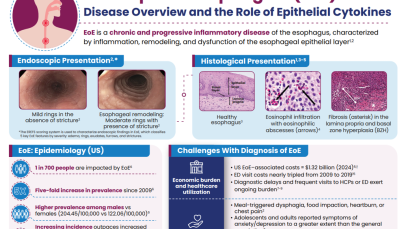
The incidence of EoE has increased rapidly in the USA in recent years.14 A recent study determined updated estimates of the prevalence and medical costs associated with EoE in the USA, using two large national insurance claim databases (MarketScan and Medicare).14 Healthcare utilization, including medications and endoscopic procedures, was quantified, and annual EoE-associated costs were estimated. Overall, 20,435 EoE cases in MarketScan (2022) and 1,913 EoE cases in Medicare (2017) were identified. Between 2009 and 2022, prevalence of EoE increased more than five-fold in the USA, from 28 cases/100,000 people to 163 cases/100,000 people.14 The economic impact is substantial, with total EoE-associated healthcare costs in the USA estimated to be $1.32 billion in 2024, when adjusted for inflation.14

Several risk factors contribute to EoE pathogenesis

Ingested food allergens (including cow’s milk, wheat, soy, and eggs) are known triggers of epithelial dysfunction and T2 inflammation in EoE, and are considered to be risk factors and drivers of the disease.4,10,15 A disrupted epithelial barrier permits allergen penetration and subsequent immune activation.10 Although food allergens are known triggers of EoE, the mechanisms behind allergen-specific immune activation are not fully understood and further research is needed. Studies would suggest that the removal of specific foods, either by dietary elimination or use of hypoallergenic formulas, can lead to disease remission.15

The involvement of aeroallergens, infectious disease, and microbiome-altering factors in infancy (including antibiotic use during the first year of life, Cesarean delivery and pre-term birth, use of acid suppressants, or a stay in a neonatal intensive care unit), have been also been implicated in several studies.15,16 Although these factors are being investigated for a possible role in EoE, they have not yet been defined as proven risk factors for EoE.16

EoE is over five-times more likely to occur in people with atopy, and the more atopic comorbidities a patient has, the more likely they are to have EoE.17,18 Much like EoE, atopic diseases such as asthma, feature a disrupted epithelial barrier, which initiates and drives long-term T2 inflammation leading to clinical symptoms.4,19 Previous research suggests that between 60% and 80% of patients have concomitant allergic conditions, including IgE-mediated food allergies, asthma, atopic dermatitis, and allergic rhinitis.4 This highlights the importance of taking a detailed medical history when evaluating the patient in order to make the correct diagnosis and decisions about next steps for care.

The importance of the epithelium and its interaction with the environment in disease pathogenesis has also been seen in children with EoE. A recent study assessed concurrent skin and esophageal dysfunction by evaluating ceramide levels in the skin of pediatric patients with EoE. Skin lipid composition of pediatric patients with EoE, but without atopic dermatitis (n=21), were compared with non-atopic dermatitis, non-EoE controls (n=17). It was found that the skin of patients with EoE have significant deficits in ceramide levels, particularly ultralong-chain fatty acid-containing ceramides, compared with the controls.20 These deficits may impair the skin’s barrier function and its interaction with environmental factors, such as allergens and microbes, supporting the concept that unified epithelial barrier dysfunction may contribute to both skin and esophageal atopic diseases.20

Learn more about epithelium-driven diseases, including asthma and chronic rhinosinusitis

Although environment is believed to play a role in EoE, genetic factors, with familial associations, are also associated with the risk of developing EoE.13,21–23 Studies have found that adult first-degree relatives of patients with EoE are at risk for EoE, particularly those who are younger, male, and have a history of atopic symptoms or diseases.18,22,23 The prevalence of EoE in first‑degree relatives ranges from 1.8% to 14.6%,22,23 and the risk of developing EoE for siblings was 2.4%, increasing to 22% for dizygotic twins.22

Several studies, including candidate-gene identification and genome-wide association studies, have identified multiple genes, including TSLP and CAPN14, that may contribute to the development of EoE.13,21,24 The overexpression of these genes have been found to disrupt the esophageal epithelial barrier and enhance immune-mediated T2 inflammation.13,21,24

Genomic analyses have identified polymorphisms in TSLP and TSLP receptor (TSLPR) associated with EoE.25–27 Single nucleotide polymorphisms residing in the TSLP gene were found to augment T2 responses, and play an important role in promoting T helper type 2 (Th2) cell differentiation, an immune cell contributing to esophageal inflammation.21,24

CAPN14 encodes an IL-13 induced, esophagus-specific proteolytic enzyme involved in epithelial homeostasis and repair, and may account for the tissue specificity of esophageal disease in EoE.21,24

The overexpression of several other genes have been found to disrupt the esophageal barrier and contribute to the development of EoE, including EMSY, STAT6, LRRC32, ANKRD27, CCL267, PDCD5, TGFB, and HIF1α.7,10,21,24,28

Chronic esophageal inflammation is associated with long-term complications in EoE

Over time, an inflammatory phenotype in EoE, primarily seen in children, may progress to a fibrostenotic or mixed inflammatory-fibrostenotic phenotype, typically seen in adults.29,30

Inflammatory phenotypes are associated with the early course of the disease and are commonly seen with children.29,30 Features of the inflammatory phenotype include: furrows, plaques, white exudates, and a normal-presenting esophagus with no evidence of fibrostenotic changes.29,30 Mixed and fibrostenotic phenotypes are associated with disease progression and the subsequent associated symptoms:4,29,31,32

  • Esophageal fibrostenosis and reduced distensibility, requiring dilation
  • Food impaction requiring endoscopic bolus removal
  • Esophageal perforation

Increasing fibrostenosis over time suggests that the natural history of EoE is one that progresses from an inflammatory to a fibrostenotic phenotype in the majority of patients.29 A retrospective Swiss study (N=200), found that in patients with untreated EoE, the longer the duration of untreated symptoms, the higher the risk of strictures (untreated EoE for 0-2 years, 17% strictures; >8-11 years, 38%; >20 years, 71% stricture rate).33 Therefore, it may be hypothesized that treatment of chronic T2 inflammation could prevent disease progression.29 Understanding the pathogenesis of EoE and the early recognition of phenotypes may also aid physicians when making treatment decisions and improve outcome assessments.

Patients with EoE often experience years of diagnostic delay.34,35 In a US study (N=677) conducted between 2000 and 2014, the average duration of symptoms of EoE in adults prior to diagnosis was estimated to be up to 10.2 years.34 This is particularly concerning, because gaps in care are associated with an increased risk of developing esophageal stricture (after a 2-year gap in care, each additional year has been associated with a 26% increased risk).36

Reasons for diagnostic delay include:

  • Patients presenting with non-specific symptoms37
  • Patients using adaptive eating behaviors that can mask their symptoms4,37
  • Lack of clinical suspicion or histological misinterpretation38
  • Lack of access to healthcare, issues with hidden costs such as travel to specialists or lacking necessary health insurance, when required39,40
  • Delayed endoscopy requests or appointments38,40
  • Lack of biopsies taken at the time of food impaction4

Increasing EoE symptom severity is associated with an increased burden on patients

Increased EoE symptom severity is associated with impaired quality of life, which includes social and emotional impact that can be worsened by strict limitations to diet/eating.3,41,42 In the USA, adolescents and adults with EoE reported symptoms of anxiety and depression to a greater extent than their healthy peers, and 75% of adults with EoE feel embarrassed by their condition.43,44 Reduced attendance at school and work is also common, with 20% of adults with EoE and 46% of caregivers reporting that they had to stop working altogether.43–45 Medical resource utilization is high for patients with EoE due to diagnostic delays and frequent visits to healthcare professionals or emergency departments.4,14,42 The average cost of an EoE-associated emergency department visit nearly tripled in the USA between 2009 and 2019 (from ~$3,700 to ~$9,000).46 The economic burden of EoE is likely to continue to rise as disease incidence and chronicity increases.42

Pathophysiology: the central role of the epithelium in homeostasis and disease

The healthy esophageal epithelium orchestrates homeostatic and pathogenic immune responses;6 intercellular junctional complexes (eg tight junctions) control paracellular transport and the basal zone of epithelium occupies less than 15% of total epithelial thickness.6,7

In EoE the esophageal epithelium is disrupted. This is characterized by:

  • Loss of structural proteins: E-cadherin, claudins, and desmoglein-1, and an imbalance between increased protease expression (CAPN14) and decreased protease inhibitor expression (SPINK7) weakens tight junctions and adherens junctions, facilitating allergen penetration and immune activation10,47,48
  • Disrupted tight junctions: apical junction complex defects and dilated intercellular spaces are observed6,7
  • Basal zone hyperplasia: the basal cell layer occupies more than 15% of the total epithelial thickness49,50
  • Subepithelial fibrosis and smooth muscle hypertrophy can also be seen6,7

Image
Esophageal inflammation
The esophageal epithelium is disrupted in EoE
Download resource

In EoE, the disrupted esophageal epithelium interacts with environmental insults.6,10 Upon allergen exposure, epithelial cells release TSLP, IL-33, and IL-25, which activate Th2 cells (via dendritic cells) and type 2 innate lymphoid cells (ILC2s). These cells, in turn, secrete T2-polarizing cytokines, including IL-4, IL-5, and IL-13, amplifying the inflammatory cascade, and contributing further to impaired epithelial barrier function.6,10,51 The chronic T2 inflammation may lead to fibrosis and remodeling, including strictures.7,10

Pathophysiology: the role of epithelial cytokines in EoE

Esophageal biopsies from patients with EoE show significantly higher epithelial-derived cytokine expression than healthy controls.1,2,52 Variants in the genes encoding the alarmin TSLP are associated with increased risk of developing EoE, with TSLP expression found to be increased in patients with EoE.7 The overexpression of several critical genes, including genes encoding TSLP, may disrupt the esophageal barrier and enhance immune-mediated inflammation.24

Increased production of antimicrobial peptides and epithelial cytokines, known to drive Th2 inflammation and eosinophil activation, were also observed.2 When comparing active EoE with normal esophageal samples, all three epithelial cytokines (TSLP, IL-33, and Il-25) showed increased expression.2

Image
Epithelial cytokines are overexpressed in patients with EoE
Download resource

In a separate biopsy study, TSLP responsive-CD4+ T cells were shown to be increased in patients with EoE compared with non-EoE controls.53 TSLP responsive CD4+ T cells (ie those expressing a receptor for TSLP) had augmented production of IL-4 or IL-13 compared with non-TSLP responsive-CD4+ T cells.53 In addition, TSLP was found to induce the expression of TSLPR, providing evidence of a feed-forward loop and a driver of T2 inflammation.53

Elevated IL-33 messenger RNA expression has also been observed in pediatric patients with confirmed EoE when compared with pediatric patients without EoE.54 Analysis of biopsy samples from patients with EoE showed that the IL‑33 receptor, ST2, has been detected on esophageal-infiltrating eosinophils from patients with active EoE.55

Tumor necrosis factor-like ligand 1A (TL1A) is a newly discovered epithelial cytokine.56 Principally, TL1A has been found to synergize with IL-15 to induce several cytokines involved in the proinflammatory response,57 and has been implicated in inflammatory diseases of the gut;56 however, the role of TL1A in EoE remains poorly characterized.56

Watch this video to learn more about the pathological mechanisms that drive EoE

Evidence from EoE murine models has demonstrated that the epithelial cytokines (TSLP, IL-25, and IL-33) are elevated in EoE and play an important role in the symptomology and pathophysiology of EoE.58–60 EoE murine models have also indicated environmental triggers that may contribute to EoE pathogenesis, including, but not limited to, aeroallergens, food allergens and infectious diseases.7,61 Interestingly, a study by Doyle et al. also demonstrated that exposure to detergents, such as sodium dodecyl sulfate (SDS), found in common household products (eg toothpaste) decreases esophageal barrier integrity, stimulates IL-33 production, and promotes epithelial hyperplasia and tissue eosinophilia in murine models (exposed to 0.5% SDS for 14 days).61 These animal models, therefore, support the important role of impaired epithelial health (ie epithelial barrier dysfunction, and subsequent immune cell recruitment and activation) in EoE.58–60

There are challenges with the current management of EoE

Based on the current American College of Gastroenterology guidelines, the level of evidence to support the use of many available therapies in patients with EoE is generally assessed to be low to moderate and there is a need for additional studies, including placebo-controlled studies, to assess the efficacy of therapies in EoE.3,4 There is currently no consensus on the recommended first-line therapy for EoE; however, there are several therapeutic options for patients with EoE.4

  • Swallowed topical corticosteroids (STCs), proton pump inhibitors (PPIs), and dietary elimination are all recommended treatments3,4
    • Feeding therapy can be used adjunctively in children with a food aversion or feeding dysfunction4
  • Biologic therapy is currently considered to be a step-up therapy in difficult-to-treat patients or patients with multiple atopic conditions4
  • Esophageal dilation is used as an adjunct to medical therapy, as a treatment for esophageal strictures causing dysphagia4

The decision of which treatment to use should be individualized based on disease characteristics and patient preference, and guided using a shared decision-making framework to promote adherence and optimize clinical outcomes.4,5

The current challenges with existing therapies include suboptimal dosing regimens and the accumulation of treatment-related side effects (e.g. with corticosteroids), which may compromise patient adherence and clinical benefit.5 Although treatment with PPIs and STCs can be effective in some patients, response rates vary, with approximately two-thirds and one-third of patients, respectively, not achieving histological remission.5 Furthermore, wider issues, such as loss of response over time and lack of predictors of response, can all pose challenges with conventional pharmacological therapies.4,5,39,62

Dietary elimination is one of the recommended long-term options for patients with EoE. However, issues with response and adherence are reported.4,5 Studies have shown that half of patients do not maintain long-term diets and 30–65% of patients do not find dietary elimination to be effective (ranging from a one-food to six-food elimination diet).4 Repeated endoscopies, close monitoring and input from a dietician are required to support dietary elimination.4 A possible solution is to offer ‘diet holidays’, when patients can take a break from the diet for a set period of time, and bridge the break with medication.4

Additional research is needed to guide treatment decision-making when selecting first-line therapies for patients with different phenotypes.5 Shared decision-making with patients is key to ensuring optimum adherence and clinical outcomes.5

Despite recent advances in the diagnosis and management of EoE, challenges remain.5 Because EoE pathophysiology is characterized by disruption of the esophageal epithelium and subsequent chronic inflammation, future strategies should focus on addressing the underlying mechanisms and restoring epithelial integrity. Such approaches may help reduce inflammation, disease progression, and esophageal remodeling, which remain priorities in EoE management.5

The content for this module was created with the support of Professor Seema Aceves, Professor Arjan Bredenoord, and Professor Evan Dellon.

RELATED RESOURCES

Like this page? We have a host of other resources available in the ‘Scientific and Resource Library’ where you can find out more.

1. Travers J, et al. Sci Rep. 2017;7:17563; 2. Simon D, et al. Allergy. 2015;70:443–452; 3. Lucendo AJ, et al. United European Gastroenterol J. 2017;5:335–358; 4. Dellon ES, et al. Am J Gastroenterol. 2025;120:31–59; 5. Bredenoord AJ, et al. Am J Gastroenterol. 2022;117:1231–1241; 6. Canani BR, et al. Allergy. 2024;79:1485–1500; 7. Laky K, Frischmeyer-Guerrerio PA. J Allergy Clin Immunol. 2024;153:1485–1499; 8. de Bortoli N, et al. Dig Liver Dis. 2024;56:951–963; 9. American Academy of Pediatrics. Diagnosis of eosinophilic esophagitis. 2023. Accessed March 2, 2026. Available from: https://www.aap.org/en/patient-care/eosinophilic-esophagitis/diagnosis-of-eosinophilic-esophagitis/?srsltid=AfmBOorPaMCbTZjQ_PagA339jwUq-5nLjl3g0RWIcrxEJtOSIyqgnMR_; 10. Farah A, et al. Diagnostics (Basel). 2025;15:240; 11. Canakis A, Dellon ES. Dis Esophagus. 2025;38:doaf045; 12. Collins MH, et al. Dis Esophagus. 2017;30:1–8; 13. Dhar A, et al. Gut. 2022;71:1459–1487; 14. Thel HL, et al. Clin Gastroenterol Hepatol. 2025;23:272–280; 15. Dellon ES, Hirano I. Gastroenterology. 2018;154:319–332.e8; 16. Jensen ET, Dellon ES. J Allergy Clin Immunol. 2018;142:32–40; 17. Weir AA, et al. Ann Allergy Asthma Immunol. 2025;134:362–364; 18. Hill DA, et al. J Allergy Clin Immunol Pract. 2018;6:1528–1533; 19. Akdis CA. Nat Rev Immunol. 2021;21:739–751; 20. Goleva E, et al. J Allergy Clin Immunol. 2025;155:2075–2079.e1; 21. O’Shea KM, et al. Gastroenterology. 2018;154:333–345; 22. Alexander ES, et al. J Allergy Clin Immunol. 2014;134:1084–1092.e1; 23. Peterson K, et al. Clin Gastroenterol Hepatol. 2022;20:e957–e963; 24. Ryu S, et al. Int J Mol Sci. 2020;21:7253; 25. Rothenberg ME, et al. Nat Genet. 2010;42:289–291; 26. Sherrill JD, et al. J Allergy Clin Immunol. 2010;126:160–165.e3; 27. Kottyan LC, et al. Nat Genet. 2014;46:895–900; 28. Masterson JC, et al. J Clin Invest. 2019;129:3224–3235; 29. Dellon ES, et al. Gastrointest Endosc. 2014;79:577–585; 30. Warners MJ, et al. Am J Gastroenterol. 2018;113:836–844; 31. Chen JW, et al. Endoscopy. 2016;48:794–801; 32. Runge TM, et al. J Clin Gastroenterol. 2017;51:805–813; 33. Schoepfer AM, et al. Gastroenterology. 2013;145:1230–1236.e1-2; 34. Reed CC, et al. Clin Gastroenterol Hepatol. 2018;16:1667–1669; 35. Navarro P, et al. United European Gastroenterol J. 2022;10:507–517; 36. Chang NC, et al. Clin Gastroenterol Hepatol. 2022;20:1701-1708.e2; 37. Muir AB, et al. Clin Exp Gastroenterol. 2019;12:391–399; 38. Kanakala V, et al. Eur J Gastroenterol Hepatol. 2010;22:848–855; 39. Dorrell R, et al. Gastroenterology. 2020;158(Suppl. 1). Abstract S828; 40. Dellon ES. Ann Allergy Asthma Immunol. 2019;123:166–172; 41. Taft TH, et al. J Asthma Allergy. 2019;12:389–399; 42. Mukkada V, et al. Clin Gastroenterol Hepatol. 2018;16:495-503.e8; 43. Gold BD, et al. Gastro Hep Adv. 2024;3:1087–1097; 44. Pokrzywinski RM, et al. Adv Ther. 2020;37:4458–4478; 45. Votto M, et al. Minerva Pediatr. 2020;72:424–432; 46. Lam AY, et al. Clin Gastroenterol Hepatol. 2023;21:3041-3050.e3; 47. Azouz NP, et al. Sci Transl Med. 2018;10:eaap9736; 48. Davis BP, et al. JCI Insight. 2016;1:e86355; 49. Collins MH. Gastrointest Endosc Clin N Am. 2008;18:59–71; viii–ix; 50. Collins MH. Dig Dis. 2014;32:68–73; 51. Underwood B, et al. Ann Allergy Asthma Immunol. 2023;130:28–39; 52. Chandramouleeswaran PM, et al. PLoS One. 2016;11:e0150968; 53. Rochman Y, et al. Sci Signal. 2023;16:eadg6360; 54. Judd LM, et al. Am J Physiol Gastrointest Liver Physiol. 2016;310:G13–G25; 55. Uchida AM, et al. Allergy. 2022;77:656–660; 56. Varricchi G, et al. J Allergy Clin Immunol. 2025;155:1420–1434; 57. Holmkvist P, et al. Mucosal Immunol. 2015;8:545–558; 58. Collison AM, et al. J Allergy Clin Immunol. 2015;136:971–982; 59. Dsilva A, et al. Allergy. 2025;80:3095–3107; 60. Masuda MY, et al. J Allergy Clin Immunol. 2024;153:1355–1368; 61. Doyle AD, et al. Allergy. 2023;78:192–201; 62. Haasnoot ML, et al. Am J Gastroenterol. 2022;117:1412–1418.